Cart
0
You may also like



If a clinician has mentioned a "grade 2 ulcer" or written "Wagner 1" in your notes, you are probably left wondering what the number means and how worried you should be. Staging systems for diabetic foot ulcers exist to give the care team a shared language for depth, tissue involvement and infection — and once you understand the logic, the notes become much easier to follow.
This article explains the two staging systems you are most likely to encounter, what each grade means in plain terms, and what tends to happen at each stage. It is a reference for understanding, not a substitute for assessment. A foot ulcer is always a reason to be seen by a healthcare professional, and quickly.
Two complications usually combine. The first is peripheral neuropathy — nerve damage that reduces sensation, so a blister, a stone in the shoe or a seam rubbing may not register as pain. The second is reduced circulation, which slows the delivery of oxygen and immune cells to the skin and makes healing take longer.
As the National Institute of Diabetes and Digestive and Kidney Diseases explains, that combination is why a minor injury in a person with diabetes can become a significant wound: the damage is not felt when it starts, and the body's repair process is working at a disadvantage. Changes in foot shape, calluses over pressure points, and dry cracked skin add further starting points.
The practical consequence is that ulcers are usually found by looking, not by feeling. This is why the American Diabetes Association puts a daily visual check at the centre of foot care, including the soles and between the toes with a mirror or a second pair of eyes. Our walkthrough of a 60-second daily diabetic foot check covers exactly what to look at and in what order.
The Wagner system, published in 1981, is the classification most people encounter. It grades an ulcer primarily by depth and by the presence of infection or gangrene, on a scale of 0 to 5. It is widely used because it is quick and consistent between clinicians, and it is also widely criticised because it captures depth well but says little about circulation or infection severity. A detailed clinical description sits in the StatPearls review of diabetic foot ulceration on the NIH's National Library of Medicine site.
The skin is intact. There is no open wound. What earns the grade is the presence of risk features: a bony deformity such as a bunion, hammertoe or Charcot change, a callus over a pressure point, a previously healed ulcer, or documented loss of protective sensation.
This is the stage where the most can be done. Care at grade 0 is usually about offloading pressure, appropriate footwear, professional callus management, and regular checks. It is also the stage where footwear and sock choices genuinely matter — seams, tight cuffs and rough interior surfaces are all avoidable sources of friction on skin that cannot report pain.
An open wound involving the epidermis and possibly the dermis, but not extending through the full thickness of the skin into deeper structures. It often appears at a pressure point — the ball of the foot, the tip or top of a toe, the heel — and may be surrounded by a rim of thickened callus.
A grade 1 ulcer may look small and unremarkable, which is exactly why it gets underestimated. Typical management involves cleaning and dressing the wound, removing surrounding callus, and above all taking pressure off the area, since a wound that is walked on all day is being reinjured continuously.
The wound extends through the skin into deeper tissue — fat, tendon, ligament or joint capsule — without abscess formation or bone involvement. The clinician may be able to probe the wound and find that it goes further than the surface suggests.
Care at this stage is more intensive: regular debridement, careful wound dressing, close monitoring for infection, and strict offloading, often with a removable boot or total contact cast. Imaging is sometimes used to establish whether deeper structures are involved.
The wound now involves a deep abscess, an infected joint, or infection of the bone (osteomyelitis). Signs may include increasing pain where sensation is preserved, warmth, swelling, discharge, odour, and systemic signs such as fever or elevated blood glucose that is harder to control than usual.
This stage generally requires urgent specialist assessment, imaging, tissue or bone sampling, and prolonged antibiotic therapy. Surgical drainage or removal of infected bone is sometimes necessary.
Tissue in part of the foot — commonly toes or the forefoot — has died, usually because blood supply to that area has failed. The affected tissue may appear black, dark brown or leathery, with a demarcation line between living and dead tissue.
Management is a vascular question first: whether circulation can be restored to the area, through angioplasty or bypass, and what tissue must be removed. This is emergency territory.
Gangrene involves the whole foot or a major part of it. At this stage the priority shifts to controlling infection and preserving life and function, and major amputation is often unavoidable.
It is worth stating plainly why these last grades exist in the scale: the NIDDK notes that ulcers that do not improve with treatment, and gangrene, are what lead to amputation of a toe, a foot, or part of a leg. The reason clinicians press so hard on daily checks and early presentation is that the distance between grade 1 and grade 3 can be a matter of weeks.

Many wound clinics use the University of Texas (UT) classification alongside or instead of Wagner, because it grades two things at once. Depth runs along one axis and complicating factors along the other.
Each grade is then given a letter:
So a "IIB" is a wound reaching tendon or capsule with infection but adequate blood flow. The advantage over Wagner is immediately visible: two ulcers of identical depth can have very different outlooks depending on whether the blood supply is intact, and the UT system records that difference explicitly.
Descriptions in a table are useful, but the practical question is what should prompt a call. The following signs warrant contacting your healthcare team promptly rather than waiting for a scheduled appointment:
A useful habit: look at your socks when you take them off. Staining is often the first evidence of a wound on a foot with reduced sensation, which is one practical argument for light-coloured socks and for changing them daily.
Nothing in this section replaces your care plan. These are the routine measures that clinical guidance consistently emphasises.
Socks are a small part of a much larger picture — but they are the part that is in contact with the skin all day, and choosing badly adds friction to a foot that has no early-warning system. For the wider context, see our complete guide to diabetic socks, and if numbness is part of your picture, what to look for in socks for neuropathy.
People understandably want a number. There isn't a reliable one, because healing depends on depth, blood supply, infection, offloading and glucose control — and the same grade of ulcer can behave very differently in two people.
What is consistent is which factors move the timeline:
One more thing worth knowing: a healed ulcer site remains a vulnerable site. Recurrence rates after a first foot ulcer are high, which is why follow-up, protective footwear and continued daily checks are recommended long after the wound has closed. Changes in foot shape and comfort over time mean the footwear that suited you five years ago may not suit you now.
The Wagner classification uses six grades: grade 0 is intact skin on an at-risk foot, grade 1 a superficial ulcer, grade 2 a deeper full-thickness wound reaching tendon or capsule, grade 3 a deep abscess or bone infection, grade 4 partial gangrene of the forefoot, and grade 5 extensive gangrene of the foot. Many clinics also use the University of Texas system, which grades depth and separately records whether infection or reduced blood supply is present.
Often unremarkable — a small open area, sometimes with a rim of hard callus around it, at a pressure point like the ball of the foot, a toe, or the heel. It may be painless if sensation is reduced, and the first sign many people notice is a stain on the sock. Any new break in the skin on the foot should be assessed rather than watched.
Foot ulcers in diabetes should always be assessed by a healthcare professional rather than managed at home. Healing depends on depth, blood supply, infection and how well pressure is kept off the area, and wounds that appear minor can involve deeper tissue than the surface shows. Early professional care is associated with better outcomes.
There is no single answer. A superficial ulcer with good circulation and effective offloading may close in a matter of weeks, while a deeper or infected wound, or one where blood supply is limited, can take many months and may need vascular or surgical intervention. Your wound care team can give you a realistic expectation for your specific ulcer.
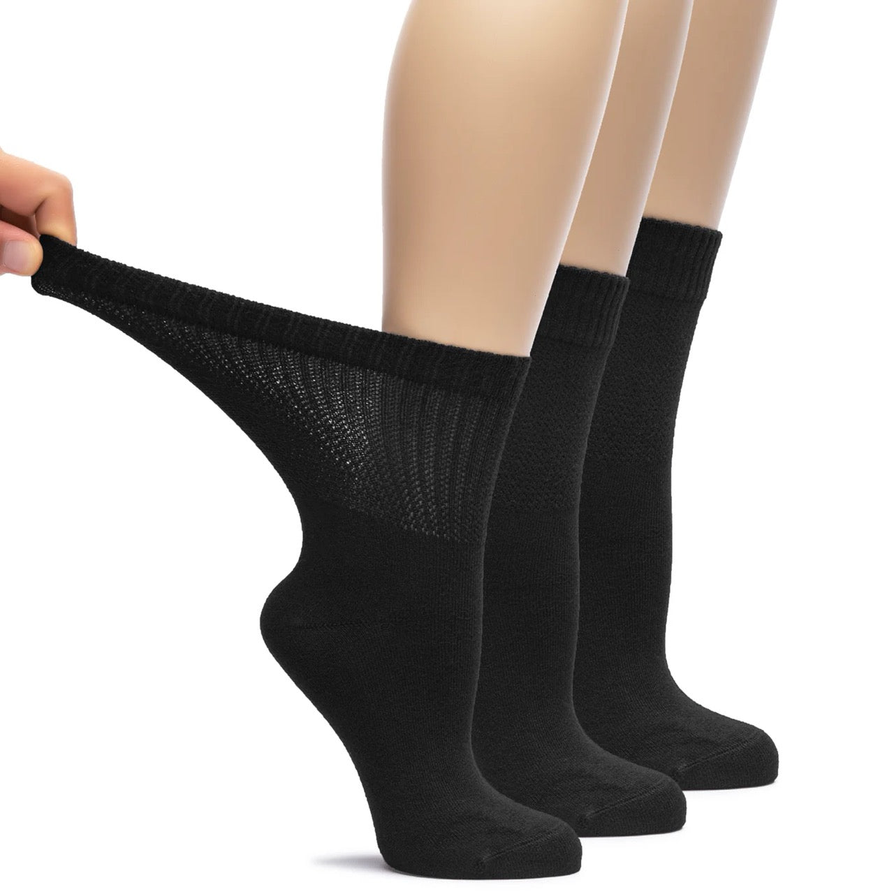
Diabetic socks are not a treatment for an ulcer and should never replace wound care. What they are designed to do is remove common sources of friction and pressure — a flat seamless toe, a wide non-binding cuff, and a smooth interior — on skin that may not be able to signal irritation. Many people find them more comfortable day to day, and light-coloured socks also make it easier to spot drainage early. Follow your care team's specific footwear guidance.
This article is for general information and is not medical advice. Talk to your healthcare provider about your own feet.
He has been working for more than 20 years in the textile industry and has work experience in many parts of the world.